
Intro
Just a few weeks ago Western Europe and the Americas suffered through one of the worst heatwaves on record [§ Copernicus 2026, § Copernicus 2026-2, § NCEI 2026, § NCEI 2026-2].
Like many others, I too went to make the best of a bad situation and spent some quality time outside; preferably close to water.
But then I saw images of a dermatologist conference on Hawaii circling around. The people knowing a damn lot about skin were sitting at the pool covered head to toe. And I started wondering, do they know something I don’t?
After all, everybody knows that you should be careful being out in intense sunlight, otherwise you will get a sunburn. Clear and painful warning that made its point; I learned my lesson, will be more careful in the future and use sunscreen. That solves it, right?
And aren’t those sunbeams anyway a generous gift, essential for life on earth as we know it.
We too rely on them, like an orphan on the feeding hand, as our primary source for vitamin D [§ Johnson 2016, § SAoN 2016, § Woodward 2016].
Why then be so careful?
So, I started to research the topic a bit and what I uncovered was anything but comforting.
As it turns out, those same sunrays, that so many love and bask themselves in, strike us with an Epic Fury that would make the american military blush.
In this article we will therefore look into…
…how bad those sunrays really are.
The mechanism of killer beams
Sunrays and UVR
Let’s first take a look at some basics. What are are we dealing with here?
Sunrays are streams of tiny packets of energy called photons, that race from the Sun through space and reach earth about eight minutes later after a journey of more than 152.1 million kilometers (94.5 million miles) [§ Espenak 2012].
But not all of those photons are the same. They posses different wavelengths and amounts of energy.
Through our eyes, we can only see a certain range of wavelengths as visible light. A little bummer, but the radiation that is most important for the scope of this article sits just beyond that visible spectrum, past violet. This form of radiation is therefore called ultraviolet or UVR and is in turn divided into the following:
| Radiation | Example wavelength | Energy/photon |
|---|---|---|
| UVA | 365 nm | ~3.40 eV |
| UVB | 300 nm | ~4.13 eV |
| UVC | 250 nm | ~4.96 eV |
UVC gets blocked by the atmosphere, but the rest gets through to us [§ D’Orazio 2013].
And just like X-rays, they are absolutely invisible to our naked eyes. Which is somewhat unfortunate considering what UVR does to you.
The UVR-damage-chain
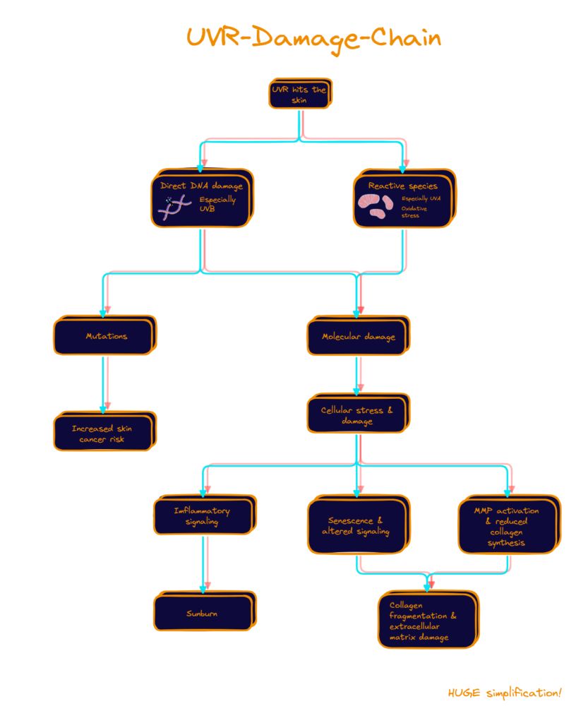
When UVR hits your skin, much of UVB is absorbed in the outer skin layer—the epidermis, our first protective layer against the oxygen rich environment we live in. UVA penetrates deeper, reaching into the dermis below. This region is a reservoir of collagen and elastin, which provide strength, structure and elasticity so that our skin can act as a cushion against all sorts of stress [§ Andrews 1990, § D’Orazio 2013, § Chambers 2020].
But neither UVA nor UVB drop by just for a quick check-in.
As energy particle-wave-thingies tend to be, those two are also always happy to make new acquaintances and spark reactions.
UVB especially loves DNA. Yes, that same DNA that is both library and instruction manual for most cells in our bodies [§ Alberts 2022].
When DNA absorbs UV energy, that energy can trigger chemical reactions between neighboring bases, creating lesions and joining molecular building blocks that most certainly do not ask to be joined together [§ Ravanat 2001, § Sinha 2002, § Cadet 2005].
UVA tends to be less direct when it comes to causing damage. Instead of being strongly absorbed by DNA itself, it can excite other molecules present in the skin. These excited molecules in turn can happily react with willing participants, generate reactive species or find other destructive outlets [§ Brem 2017].
At this stage, for one, the chain turns into a whole bouquet of damaging options, that contains classics like damaged mitochondria, inflammatory responses, or again DNA damage. And two it can happily continue even long after UV-exposure has passed [§ Premi 2015, § Fitsiou 2021, § Salminen 2022, § Yan 2025].
Repair forces
Fortunately, our cells are not entirely helpless.
They have systems that detect and repair damage. Depending on what happened and how severe it was, a damaged cell may repair itself and continue working. Or the damage can be so severe that it has to alter its behavior, stop dividing, or, if things have gone particularly badly, destroy itself [§ Matt 2016].
And these responses come with consequences of their own.
Damaged acells activate signaling pathways and release inflammatory signal molecules (e. g. cytokines and prostaglandins). These molecules also nudge other cells to join in and amplify the inflammatory response [§ Kammeyer 2015.
Under normal circumstances, inflammation is primarily a defense and repair response. It helps remove damaged material, coordinate immune activity and support tissue repair [§ Chen 2010]. But if the inflammatory response becomes too strong or persists for too long, the same signals that help coordinate repair can begin to disrupt normal function.
Chronic inflammation can promote oxidative stress, impair regeneration and contribute to the breakdown of the surrounding extracellular matrix.
Inflammation can thereby contribute to other long-term consequence of UV-induced damage.
In the case of senescence, cells enter a state in which they stop dividing but remain metabolically active [§ Fitsiou 2021].
And these senescent cells can develop what is called a senescence-associated secretory phenotype (SASP). In this state, they release cytokines, growth factors and matrix-degrading enzymes that further promote inflammation [§ Fitsiou 2021].
In this way, UV-induced damage can contribute not only to acute inflammation, but also to a longer-lasting environment of inflammation, impaired regeneration and extracellular-matrix degradation.
Meanwhile in the dermis, UV exposure activates another particularly relevant group of molecules: matrix metalloproteinases, or MMPs [§ Fisher 1998].
These MMPs are involved in the remodeling the extracellular matrix, or what keeps cells stable and in form. Two major components of that matrix are collagen, which gives skin much of its tensile strength, and elastin, which helps it return to shape. Together with the rest of the extracellular matrix, collagen and elastin provide the structural scaffolding that keeps skin strong and resilient [§ Fisher 1997, § Fisher 1998].
Under UVR, these MMPs receive so many work requests that their activity scales up so much that it contributes to the breakdown and fragmentation of collagen. At the same time, UV also reduces the production of new collagen.
So with repeated exposure, the supporting matrix of the skin, that keeps it up and firm, becomes increasingly fragmented and disorganized. And as that structural support deteriorates, the macroscopic signs of photoaging begin to appear: wrinkles, reduced elasticity, altered pigmentation, rougher texture and loss of skin tone and resilience [§ Fisher 1997, § Fisher 1998, § D’Orazio 2013, § Rittie 2015].
And because of all these effects, scientists consider UVR the leading environmental driver of premature skin aging [§ Fisher 2002, § Yaar 2007, § Flament 2013, § Tsai 2026].
Ultraviolet light through sun exposure is the leading driver of premature skin aging.
Alarming, right?
Lack of detection
But even more alarming is the body’s absolute absence of a direct alarm. We don’t feel UV photons striking our skin; there is no specific heat, or some color or a weird smell. Our sensory nervous system does not provide us with a direct perception of UV intensity [§ Lopes 2016].
What we do feel is pain. The pain of the major inflammation of the affected area—also called sunburn. The problem is, that we get sunburn hours after the fact with peak redness, warmth, pain and all the other charming symptoms, around a day later [§ Brenner 2008, § Brenner 2009, § Rhodes 2009].
That is not an alarm signal, but an educational beating; at best.
Sunburn is a late, incomplete warning.
Oh, but it gets better.
Because sunburn isn’t even a required outcome in the UVR-damage-chain. Yes, you understood that right.
For sunburn to occur a sufficient amount of cells must release molecular distress signals (e. g. IL-1, IL-6 and TNF-α) that kick off the inflammation response [§ Brenner 2009, § Rhodes 2009]. That means that sunburn is only one possible downstream response of cellular damage caused by UVR [§ Seité 2010, § Shih 2018].
We can happily develop as many wrinkles and cancer nuclei as we want without ever having suffered a single day of red swollen sun-pain.
No sunburn does not mean no damage.
Conclusions
1. UVR can’t be felt
UVR ≠ heat
UVR can’t be felt directly. We don’t have sensory neurons that tell us how much ultraviolet radiation is hitting our skin. The warmth we associate with sunshine comes largely from other parts of solar radiation and from our surroundings—not from some convenient built-in UV meter.
That means substantial UV exposure can occur even when it feels perfectly pleasant outside: on a cool day, in the mountains, in winter, when there is wind or while standing in water.
Your skin may be getting bombarded while your senses are telling our: Lovely weather, mate.
We can’t feel how much UVR we are receiving.
2. Sunburn is a late warning
Sunburn does not tell you when UV damage begins.
It is a downstream inflammatory response to UV exposure. By the time your skin becomes red, warm and painful, molecular and cellular responses to UVR have already been underway for hours.
And even that warning system is incomplete.
UVR can cause molecular and cellular damage at exposures that never produce visible sunburn. So even without entering lobster mode, your skin may still have accumulated photodamage.
No sunburn does not mean no damage.
3. Don’t use your skin as a UV detector
If our senses cannot reliably tell us how much UVR we are receiving, we need something better. One of the most useful tools is the UV Index.
The UV Index is a standardized measure of UV radiation weighted according to how effectively different wavelengths produce erythema, or sunburn.
The higher the UV index, the more cautious you should be.
Don’t listen to your skin. Check the data.
4. Time matters
But the UV Index only tells you about the intensity of UV radiation. Your actual exposure also depends on how long you stay exposed.
A few minutes here and a few minutes there do not magically disappear simply because none of them produced a sunburn. Repeated UV exposure accumulates over time and contributes to long-term photodamage [§ Lavker 1995].
Judge UV by exposure, not by how the day feels.
Outro
You do not need to declare war on daylight, but if UVR is high, reduce the dose: seek shade, cover exposed skin, use sunscreen and limit unnecessary exposure.
Because your skin and you are amazing at many things. Evaluating the danger of those invisible bullets the sun shoots us with is not one of them.
Sources
| Key | Citation |
|---|---|
| § Alberts 2022 | Alberts, B., Heald, R., Johnson, A., Morgan, D., Raff, M., Roberts, K., & Walter, P. (2022). Molecular biology of the cell (7th ed.). W. W. Norton & Company |
| § Andrews 1990 | Arnold, H. L., Andrews, G. C., Odom, R. B., & James, W. D. (1990). Andrews’ diseases of the skin: clinical dermatology. (No Title). |
| § Brem 2017 | Brem, R., Guven, M., & Karran, P. (2017). Oxidatively-generated damage to DNA and proteins mediated by photosensitized UVA. Free radical biology & medicine, 107, 101–109. https://doi.org/10.1016/j.freeradbiomed.2016.10.488 |
| § Brenner 2009 | Brenner M, Hearing VJ. The protective role of melanin against UV damage in human skin. Photochem Photobiol. 2008 May-Jun;84(3):539-49. doi: 10.1111/j.1751-1097.2007.00226.x. PMID: 18435612; PMCID: PMC2671032. |
| § Brenner 2008 | Brenner, M. and Hearing, V.J. (2008), The Protective Role of Melanin Against UV Damage in Human Skin†. Photochemistry and Photobiology, 84: 539-549. https://doi.org/10.1111/j.1751-1097.2007.00226.x |
| § Cadet 2005 | Cadet, J., Sage, E., & Douki, T. (2005). Ultraviolet radiation-mediated damage to cellular DNA. Mutation Research/Fundamental and Molecular Mechanisms of Mutagenesis, 571(1–2), 3–17. https://doi.org/10.1016/j.mrfmmm.2004.09.012 |
| § Chambers 2020 | Chambers, E. S., & Vukmanovic-Stejic, M. (2020). Skin barrier immunity and ageing. Immunology, 160(2), 116–125. https://doi.org/10.1111/imm.13152 |
| § Chen 2010 | Chen, G. Y., & Nuñez, G. (2010). Sterile inflammation: sensing and reacting to damage. Nature Reviews Immunology, 10(12), 826-837. |
| § Copernicus 2026 | Copernicus Climate Change Service. (2026, August 12). Exceptionally hot and dry conditions fuel wildfires in Europe as ocean surface temperatures reach record highs for July. https://climate.copernicus.eu/exceptionally-hot-and-dry-conditions-fuel-wildfires-europe-ocean-surface-temperatures-reach-record |
| § Copernicus 2026-2 | Copernicus Climate Change Service. (2026). Surface air temperature for July 2026. https://climate.copernicus.eu/surface-air-temperature-july-2026 |
| § D’Orazio 2013 | D’Orazio, J., Jarrett, S., Amaro-Ortiz, A., & Scott, T. (2013). UV radiation and the skin. International Journal of Molecular Sciences, 14(6), 12222–12248. https://doi.org/10.3390/ijms140612222 |
| § Espenak 2012 | Espenak, F. (2012, January 6). Eclipses and the Moon’s orbit. NASA Goddard Space Flight Center. NASA – Eclipses and the Moon’s Orbit |
| § Fisher 1998 | Fisher, G. J., & Voorhees, J. J. (1998). Molecular mechanisms of photoaging and its prevention by retinoic acid: ultraviolet irradiation induces MAP kinase signal transduction cascades that induce Ap-1-regulated matrix metalloproteinases that degrade human skin in vivo. The journal of investigative dermatology. Symposium proceedings, 3(1), 61–68. |
| § Fisher 2002 | Fisher, G. J., Kang, S., Varani, J., Bata-Csorgo, Z., Wan, Y., Datta, S., & Voorhees, J. J. (2002). Mechanisms of photoaging and chronological skin aging. Archives of Dermatology, 138(11), 1462–1470. https://doi.org/10.1001/archderm.138.11.1462 |
| § Fisher 1997 | Fisher, G. J., Wang, Z. Q., Datta, S. C., Varani, J., Kang, S., & Voorhees, J. J. (1997). Pathophysiology of premature skin aging induced by ultraviolet light. The New England Journal of Medicine, 337(20), 1419–1428. https://doi.org/10.1056/NEJM199711133372003 |
| § Fitsiou 2021 | Fitsiou, E., Pulido, T., Campisi, J., Alimirah, F., & Demaria, M. (2021). Cellular Senescence and the Senescence-Associated Secretory Phenotype as Drivers of Skin Photoaging. The Journal of investigative dermatology, 141(4S), 1119–1126. https://doi.org/10.1016/j.jid.2020.09.031 |
| § Flament 2013 | Flament, F., Bazin, R., Laquieze, S., Rubert, V., Simonpietri, E., & Piot, B. (2013). Effect of the sun on visible clinical signs of aging in Caucasian skin. Clinical, cosmetic and investigational dermatology, 6, 221–232. https://doi.org/10.2147/CCID.S44686 |
| § Kammeyer 2015 | Kammeyer, A., & Luiten, R. M. (2015). Oxidation events and skin aging. Ageing research reviews, 21, 16–29. https://doi.org/10.1016/j.arr.2015.01.001 |
| § Lavker 1995 | Lavker, R. M., Gerberick, G. F., Veres, D., Irwin, C. J., & Kaidbey, K. H. (1995). Cumulative effects from repeated exposures to suberythemal doses of UVB and UVA in human skin. Journal of the American Academy of Dermatology, 32(1), 53–62. https://doi.org/10.1016/0190-9622(95)90184-1 |
| § Lopes 2016 | Lopes, D. M., & McMahon, S. B. (2016). Ultraviolet radiation on the skin: a painful experience?. CNS neuroscience & therapeutics, 22(2), 118-126. |
| § Matt 2016 | Matt, S., & Hofmann, T. G. (2016). The DNA damage-induced cell death response: a roadmap to kill cancer cells. Cellular and molecular life sciences : CMLS, 73(15), 2829–2850. https://doi.org/10.1007/s00018-016-2130-4 |
| § Johnson 2016 | Matthew P. Johnson; Photosynthesis. Essays Biochem 31 October 2016; 60 (3): 255–273. doi: https://doi.org/10.1042/EBC20160016 |
| § NCEI 2026-2 | National Centers for Environmental Information. (2026, August 10). Assessing the U.S. temperature and precipitation in July 2026. National Oceanic and Atmospheric Administration. https://www.ncei.noaa.gov/news/national-climate-202607 |
| § NCEI 2026 | National Centers for Environmental Information. (2026, August 11). Assessing the global temperature and precipitation in July 2026. National Oceanic and Atmospheric Administration. https://www.ncei.noaa.gov/news/global-climate-202607 |
| § Premi 2015 | Premi, S., Wallisch, S., Mano, C. M., Weiner, A. B., Bacchiocchi, A., Wakamatsu, K., Bechara, E. J. H., Halaban, R., Douki, T., & Brash, D. E. (2015). Chemiexcitation of melanin derivatives induces DNA photoproducts long after UV exposure. Science, 347(6224), 842–847. https://doi.org/10.1126/science.1256022 |
| § Ravanat 2001 | Ravanat, J. L., Douki, T., & Cadet, J. (2001). Direct and indirect effects of UV radiation on DNA and its components. Journal of photochemistry and photobiology. B, Biology, 63(1-3), 88–102. https://doi.org/10.1016/s1011-1344(01)00206-8 |
| § Rhodes 2009 | Rhodes, L. E., Gledhill, K., Masoodi, M., Haylett, A. K., Brownrigg, M., Thody, A. J., Tobin, D. J., & Nicolaou, A. (2009). The sunburn response in human skin is characterized by sequential eicosanoid profiles that may mediate its early and late phases. FASEB journal : official publication of the Federation of American Societies for Experimental Biology, 23(11), 3947–3956. https://doi.org/10.1096/fj.09-136077 |
| § Rittie 2015 | Rittié, L., & Fisher, G. J. (2015). Natural and sun-induced aging of human skin. Cold Spring Harbor Perspectives in Medicine, 5(1), a015370. https://doi.org/10.1101/cshperspect.a015370 |
| § Salminen 2022 | Salminen, A., Kaarniranta, K., & Kauppinen, A. (2022). Photoaging: UV radiation-induced inflammation and immunosuppression accelerate the aging process in the skin. Inflammation research : official journal of the European Histamine Research Society … [et al.], 71(7-8), 817–831. https://doi.org/10.1007/s00011-022-01598-8 |
| § SAoN 2016 | Scientific Advisory Committee on Nutrition. (2016). Vitamin D and health. GOV.UK. https://www.gov.uk/government/publications/sacn-vitamin-d-and-health-report� |
| § Seité 2010 | Seité, S., Fourtanier, A., Moyal, D., & Young, A. R. (2010). Photodamage to human skin by suberythemal exposure to solar ultraviolet radiation can be attenuated by sunscreens: a review. The British journal of dermatology, 163(5), 903–914. https://doi.org/10.1111/j.1365-2133.2010.10018.x |
| § Shih 2018 | Shih, B. B., Farrar, M. D., Cooke, M. S., Osman, J., Langton, A. K., Kift, R., Webb, A. R., Berry, J. L., Watson, R. E. B., Vail, A., de Gruijl, F. R., & Rhodes, L. E. (2018). Fractional Sunburn Threshold UVR Doses Generate Equivalent Vitamin D and DNA Damage in Skin Types I-VI but with Epidermal DNA Damage Gradient Correlated to Skin Darkness. The Journal of investigative dermatology, 138(10), 2244–2252. https://doi.org/10.1016/j.jid.2018.04.015 |
| § Sinha 2002 | Sinha, R. P., & Häder, D. P. (2002). UV-induced DNA damage and repair: a review. Photochemical & photobiological sciences : Official journal of the European Photochemistry Association and the European Society for Photobiology, 1(4), 225–236. https://doi.org/10.1039/b201230h |
| § Tsai 2026 | Tsai, J., & Kang, S. (2026). Photoaging: Update on Pathogenesis, Prevention, and Treatment. Annals of dermatology, 38(3), 167–176. https://doi.org/10.5021/ad.26.006 |
| § Woodward 2016 | Woodward W. Fischer, James Hemp, Jena E. Johnson. 2016. Evolution of Oxygenic Photosynthesis. Annual Review of Earth and Planetary Sciences 44:647-683. https://doi.org/10.1146/annurev-earth-060313-054810 |
| § Yaar 2007 | Yaar, M., & Gilchrest, B. A. (2007). Photoageing: mechanism, prevention and therapy. The British journal of dermatology, 157(5), 874–887. https://doi.org/10.1111/j.1365-2133.2007.08108.x |
| § Yan 2025 | Yuan, X., Li, H., Lee, J. S., & Lee, D. H. (2025). Role of Mitochondrial Dysfunction in UV-Induced Photoaging and Skin Cancers. Experimental dermatology, 34(5), e70114. https://doi.org/10.1111/exd.70114 |
Leave a Reply